What is the localization of central sulcus?
Central sulcus localization The central sulcus can be localized with MEG by two independent measurements, by localizing the primary sensory cortex, S1, and the primary motor cortex, M1. Stimulation of sensory nerves elicits somatosensory evoked fields (SEFs) generated at S1.
How to locate central sulcus?
Surprisingly, the most reliable way to find the central sulcus is not by inspecting the lateral surface of the brain, where this is one of the longest and deepest sulci of the human cerebral cortex. Rather, the best way to find the central sulcus is to start on the medial surface of the hemisphere.
What is the location and function of the sulci?
Sulci surround each gyrus, and together, the gyri and sulci help to increase the surface area of the cerebral cortex and form brain divisions. They form brain divisions by creating boundaries between the lobes, so these are easily identifiable and serve to divide the brain into two hemispheres.
Where is the sulcus located?
The central sulcus is between the frontal lobe and parietal lobe (2–5). Gyrus present anterior to the central sulcus is the precentral gyrus. The precentral gyrus is limited anteriorly by the precentral sulcus (Figure1A, 1C, 2A) (1, 5–7, 10).
What is the location and function of the central sulcus?
Gross anatomy The central sulcus separates the frontal lobe from the parietal lobe, and more specifically separates the primary motor cortex anteriorly from the primary somatosensory cortex posteriorly 1.
What functional area is related to central sulcus?
The central sulcus separates the frontal lobe from the parietal lobe, and more specifically separates the primary motor cortex anteriorly from the primary somatosensory cortex posteriorly 1.
How deep is the central sulcus?
Depth of the central sulcus was 1.333±0.100cm to 1.029±0.125cm in male and 1.173±0.144cm to 1.01±0.200cm in female.
What would happen if the central sulcus is damaged?
The central sulcus separates the frontal from the parietal lobe, and on each side of this sulcus lie the pre-central gyrus and the post-central gyrus. Damage to both frontal lobes may produce an alteration in personality, a loss of normal inhibitions and incontinence.
What is central sulcus in CT brain?
Central sulcus in a very important landmark that separate the frontal lobe from parietal lobe. It separate the precentral gyrus (motor cortex) from post central gyrus (sensory cortex). A few signs/ways to recognize central sulcus has been described. It is important to note that not all signs present in any one patient.
What are the functional areas of sulci and gyri?
➢ Sulci: superior & inferior ➢ Gyri: superior, middle & inferior. associated with taste, vestibular, visceral sensation, secondary somatic and auditory. Cerebral cortex is necessary for conscious awareness and thought, memory and intellect.
What is the importance of sulci?
Brain gyri and sulci serve two very important functions: They increase the surface area of the cerebral cortex and they form brain divisions. Increasing the surface area of the brain allows more neurons to be packed into the cortex so that it can process more information.
What is the function of the orbital sulci?
The orbital gyri are located on the inferior surface of the frontal lobe. There are four gyri and they are divided by the H-shaped orbital sulci. They have a role in the perception of odors.
How to know central sulcus?
The central sulcus is identified on the sagittal images and, using the lateral view of the skull as a reference image, the topographic information is transferred to the axial images.
What is the primary significance of the central sulcus?
The correct localization of the central sulcus is hugely important on cross-sectional imaging as it defines the primary motor cortex anteriorly and the primary sensorimotor cortex posteriorly.
What is the difference between the central sulcus and the central gyrus?
The surface of the brain is folded with each crest being termed a gyrus and each groove between them a sulcus. The central sulcus separates the frontal from the parietal lobe and on each side of this sulcus lie the pre-central gyrus (in front) and the post-central gyrus (behind).
Where is the sulcus located in the body?
The central sulcus (the sulcus of Rolando) forms the boundary between the frontal and the parietal lobes on the lateral and medial surfaces of the cerebral hemispheres (Figs.
What is sulcus and its function?
Sulci are grooves on the brain’s surface. They are situated between the gyri. They divide the cerebral cortex into gyri. The sulcus that divides the left and right brain hemispheres is the medial longitudinal fissure.
Where is the post central sulcus?
The postcentral sulcus is a sulcus of the parietal lobe that separates the postcentral gyrus from the remainder of the parietal lobe, thus dividing the primary somatosensory cortex from the secondary somatosensory cortex. It runs parallel and posterior to the central sulcus.
What are the functions of the central sulcus?
The central sulcus separates the parietal lobe (blue) and the frontal lobe (lime green). The central sulcus is a prominent landmark of the brain, separating the parietal lobe from the frontal lobe and the primary motor cortex from the primary somatosensory cortex.
What happens if the central sulcus is damaged?
Cerebral amyloid angiopathy raises the danger of stroke and dementia triggered by bleeding. In a study conducted (Chamarthy et al., 2012), the association of damage to the central sulcus was associated with cerebral amyloid angiopathy (CAA).
What is located anterior to the central sulcus?
The precentral gyrus, which is directly anterior to the central sulcus and runs parallel to it, contains the primary motor cortex (Brodmann area 4). The primary motor cortex is responsible for controlling the voluntary movements of specific body parts.
What does the central sulcus separate?
The central sulcus runs posterior-medial to anterior-lateral and separates the frontal lobe from the parietal lobe.
What causes a deep central sulcus?
This problem often occurs in horses with high, contracted heels and is a common feature in club feet. It is usually indicative of an underlying lameness-related problem rather than a cause of lameness. Thrush tends to develop in the grooves and worsens the problem by penetrating deep into the tissues.
What landmarks would you use to identify the central sulcus?
using four landmarks: (i) inverted omega of the precentral gyrus [13], (ii) the inverted T sign at the termination of the superior frontal sulcus at the precentral gyrus [14], (iii) marginal ramus of the cingular sulcus or “pli de passage frontoparietal superieur” [15] and (iv) termination of precentral gyrus behind …
What is another name for the central sulcus?
The central sulcus, also known as the sulcus of Rolando, is one of the largest sulci present in the brain. It is located between the frontal and parietal lobes.
How to recognize the central sulcus?
The lower T sign is one of the features useful in identifying the central sulcus on cross-sectional imaging. It relies on identifying the inferior frontal sulcus which intersects the precentral sulcus in a “T” junction, thus defining the precentral gyrus. The central sulcus is the next posterior sulcus.
What is the significance of the sulcus?
Sulci, the grooves, and gyri, the folds or ridges, make up the folded surface of the cerebral cortex. Larger or deeper sulci are termed fissures, and in many cases the two terms are interchangeable. The folded cortex creates a larger surface area for the brain in humans and other mammals.
What is localization of the cerebral cortex?
Your cerebral cortex is the outer layer that lies on top of your cerebrum. Your cerebrum is the largest area of your brain. Your cerebrum divides your brain into two halves called hemispheres. The hemispheres are attached by a bundle of nerve fibers called the corpus callosum.
How do you localize a CNS lesion?
A simple practical method for localizing lesions in the brain is the use of four axons, the axon of the sensomotor pathaways, the axon of the optic pathway, the axon of the cranial nerves and the axon of ventricular system (Figure 5-1, axon 1-5).
What is localization of the brain regions?
Localisation is where a specific brain area is associated directly with a particular function. Lateralisation is where the brain has two hemispheres connected by the corpus callosum that are not entirely alike. Each hemisphere of the brain is specialised to perform certain functions or processes.
What are the boundaries of the central sulcus?
The central sulcus (the sulcus of Rolando) forms the boundary between the frontal and the parietal lobes on the lateral and medial surfaces of the cerebral hemispheres (Figs.
What is the function of the central sulcus?
Where is the central sulcus located?
Where does the central sulcus terminate?
How do you find the central sulcus?
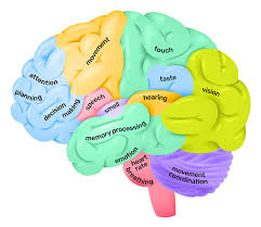
Think of it like this: the central sulcus is like a big, deep ditch running down the middle of your brain. It’s kind of like the line of scrimmage in a football game, separating the front of the brain (the frontal lobe) from the back of the brain (the parietal lobe).
Where is the central sulcus located?
You can find the central sulcus by running your fingers along the middle of your head from your forehead to the back of your skull. It’s right under your scalp, and if you could see through your skull, you’d see it there.
It’s not just a random groove, though. The central sulcus is a landmark in the brain, and it’s crucial for how your brain works.
What does the central sulcus do?
The central sulcus is important because it separates the frontal lobe and the parietal lobe of the brain.
The frontal lobe is responsible for things like planning, decision-making, and movement.
The parietal lobe is responsible for things like touch, temperature, and pain sensation.
So, the central sulcus acts like a boundary between these two important areas.
Why is the central sulcus so important?
The central sulcus is important because it allows the different parts of your brain to communicate with each other. Imagine it as a bridge between the two lobes.
The frontal lobe needs to send signals to the parietal lobe to control movement. For example, when you decide to pick up a cup of coffee, the frontal lobe sends signals through the central sulcus to the parietal lobe to tell your hand to move.
The parietal lobe also needs to send signals to the frontal lobe to tell you about your environment. For example, if you touch a hot cup of coffee, the parietal lobe sends signals through the central sulcus to the frontal lobe to tell you that the cup is hot.
So, the central sulcus is a vital communication link between the two lobes.
What happens if the central sulcus is damaged?
Damage to the central sulcus can lead to a variety of problems, depending on the severity of the damage and the specific areas affected.
* If the frontal lobe is damaged, you might have difficulty with planning, decision-making, or controlling your movements.
* If the parietal lobe is damaged, you might have difficulty with touch, temperature, or pain sensation.
How is the central sulcus studied?
We study the central sulcus using a variety of techniques, including:
Brain imaging. This allows us to see the structure of the brain, including the central sulcus, in great detail.
Electroencephalography (EEG). This technique measures the electrical activity of the brain, which can be used to study how the central sulcus functions.
Transcranial magnetic stimulation (TMS). This technique uses magnetic pulses to stimulate or inhibit the activity of specific brain areas, including the central sulcus.
By using these techniques, we can learn more about the structure and function of the central sulcus and its role in brain function.
What are some interesting facts about the central sulcus?
* The central sulcus is one of the most prominent features of the brain.
* It’s present in all mammals, including humans.
* The central sulcus is located on the precentral gyrus and the postcentral gyrus.
* The precentral gyrus is the area of the brain that controls movement.
* The postcentral gyrus is the area of the brain that receives sensory information.
FAQs
What is the central sulcus also known as?
The central sulcus is also known as the Rolandic sulcus.
What is the difference between the central sulcus and the central gyrus?
The central sulcus is the groove, and the central gyrus is the ridge on either side of it. Think of the central sulcus as a valley and the central gyrus as the hills on either side of the valley.
What are some diseases that affect the central sulcus?
Several diseases can affect the central sulcus, including:
Stroke. This can damage the central sulcus and lead to problems with movement, sensation, or both.
Alzheimer’s disease. This disease can cause atrophy of the brain, including the central sulcus.
Epilepsy. Seizures can originate from the central sulcus and cause a variety of symptoms.
Can I see the central sulcus on a brain scan?
Yes, you can see the central sulcus on a brain scan, such as an MRI or CT scan.
Why is the central sulcus important for learning?
The central sulcus plays a critical role in learning by enabling the frontal lobe to communicate with the parietal lobe, allowing for integration of sensory information and motor planning.
What is the function of the precentral gyrus and the postcentral gyrus?
The precentral gyrus is responsible for voluntary movement, while the postcentral gyrus is responsible for processing sensory information, including touch, temperature, and pain.
Is the central sulcus the same in all brains?
No, the central sulcus can vary in shape and size between individuals.
How can I protect my central sulcus?
You can protect your central sulcus by taking steps to prevent stroke, Alzheimer’s disease, and epilepsy.
I hope this information about the central sulcus was helpful! Remember, it’s a crucial part of your brain and plays a vital role in how you think, feel, and move.
See more here: How To Locate Central Sulcus? | Central Sulcus Location And Function
Central sulcus: Anatomy, location and contents | Kenhub
The central sulcus, also known as the sulcus of Rolando, is one of the largest sulci present in the brain. It is located between the frontal and parietal lobes. Apart from being the boundary between the two Kenhub
Central sulcus – Location, Function and Pictures
Central sulcus Function Its primary function is to separate the parietal lobe of the cerebral cortex from the frontal lobe. The parietal lobe also referred to as the Parietal cortex, is the section of the cerebral KnowYourBody.net
Central sulcus | Radiology Reference Article
The central sulcus separates the frontal lobe from the parietal lobe, and more specifically separates the primary motor cortex anteriorly from the primary somatosensory cortex posteriorly 1. Radiopaedia
Central Sulcus | Complete Anatomy – Elsevier
The central sulcus (aka central sulcus of cerebrum) is a groove along the surface of the cerebral cortex that separates the frontal lobe from the parietal lobe. It also separates Elsevier
Central Sulcus – Practical Psychology
The central sulcus is the central groove found in the cerebral cortex of the brain, it is also known as the Rolando Fissure. The central sulcus connects the frontal and parietal Practical Psychology
Central Sulcus – an overview | ScienceDirect Topics
A central sulcus (arrowhead) separates several anterior short gyri from one or two posterior long gyri, all of which radiate in a fan-like manner from the anteroventral part of ScienceDirect
Central sulcus – e-Anatomy – IMAIOS
Definition. The central sulcus, which is also referred to as the Rolando fissure or central fissure, is located approximately in the middle of the lateral surface of the cerebral IMAIOS
Central Sulcus – an overview | ScienceDirect Topics
Central sulcus localization. The central sulcus can be localized with MEG by two independent measurements, by localizing the primary sensory cortex, S1, and the ScienceDirect
Medical Neuroscience | Tutorial Notes – Duke University
The central sulcus is one of the most important landmarks in the human brain for clinicians and neuroscientists because it precisely divides the somatic sensory cortex of the Duke Histology
See more new information: pilgrimjournalist.com
2-Minute Neuroscience: Lobes And Landmarks Of The Brain Surface (Lateral View)
Cerebral Cortex (Function, Covering, Lobes, Sulcus, Gyrus, Fissures) | Anatomy
Gyri Of The Brain – Learn In 4 Minutes
Finding The Central Sulcus
3 Tips To Find The Central Sulcus
Link to this article: central sulcus location and function.

See more articles in the same category here: https://pilgrimjournalist.com/wiki/